Foot Muscles Mri / 52 best images about MRI anatomy on Pinterest | Head and ... : Lateral and medial processes of calcaneal tuberosity.. Explore more like foot muscle anatomy mri. Top suggestions for foot muscle anatomy mri. Muscle mri sequences & patterns asymmetric myopathy hereditary acquired connective tissue neurogenic. The muscles with proximal attachments at points outside the foot are referred to as extrinsic. Muscles of the foot muscle origin insertion nerve supply extensor digitorum brevis distal part of the lateral and superior surfaces of the calcaneus and the apex of the inferior extensor.
This article reviews the use of magnetic resonance imaging (mri) in the evaluation of the foot, including a mri of the foot. In this weeks video, we have a look at muscle edema in the intrinsic and plantar muscles of the foot and what it can mean.patreons can access original dicom. Learn about foot and ankle mri here. The muscles lie within a flat fascia on the dorsum of the foot (fascia dorsalis pedis) and are innervated by the deep fibular interestingly the dorsal foot muscles generally have no insertion at the little toe. | find, read and cite all the research you the foot arch and the foot functional capacity is strongly related to the strength of the flexor muscles.
Foot anatomy mri coronal Images from musculoskeletalkey.com Magnetic resonance imaging—mri—uses magnetic fields and radio waves to examine the internal structures of your body. Mri and ultrasound have been utilised in the assessment of the plantar intrinsic foot muscles. Magnetic resonance imaging (mri) is the method. Top suggestions for foot muscle anatomy mri. Posted by radiologyer at 8:12 am. The abductor digiti minimi muscle is on the lateral side of the foot and contributes to the large lateral plantar eminence on the sole. Gray's anatomy for students, 2nd ed. A measure of diabetic neuropathy.
Neurovascular abnormalities and skin abnormalities in the affected limb were identified on mri in 1 and 2 patients, respectively.
Posted by radiologyer at 8:12 am. The muscles acting on the foot can be divided into two distinct groups; Head, neck, arm, foot, pelvis, etc. Intrinsic foot muscles differ from extrinsic foot muscles, which have their origins in the leg and the long tendons cross the ankle joint complex 27. Top suggestions for foot muscle anatomy mri. Lateral and medial processes of calcaneal tuberosity. If muscles, tendons and bones are not in use they will. This is a 30 year old with swelling on the lateral aspect of foot with evidence of soft tissue lesion in relation to the lateral aspect of the talus which appears isointense to the muscles on t1 and t2. However, on mri images, no muscular abnormalities were detected. The extrinsic muscles of the foot originate from the anterior, posterior and lateral compartments of the leg. If you'd like to support us and get something great in return. In this weeks video, we have a look at muscle edema in the intrinsic and plantar muscles of the foot and what it can mean.patreons can access original dicom. By muhammad ali, mb bs;
Explore more like foot muscle anatomy mri. Magnetic resonance imaging (mri) is the method. Muscle mri sequences & patterns asymmetric myopathy hereditary acquired connective tissue neurogenic. Lateral and medial processes of calcaneal tuberosity. The muscles acting on the foot span from above the knee to various points on the foot skeleton.
Pin by Varsha Kunwar Gautam on MRI anatomy | Ankle anatomy ... from i.pinimg.com Feet and ankles ankle muscle anatomy of foot muscles of foot muscles foot foot muscles anatomy muscle composite video showing multiple mri images including: Head, neck, arm, foot, pelvis, etc. A measure of diabetic neuropathy. Neurovascular abnormalities and skin abnormalities in the affected limb were identified on mri in 1 and 2 patients, respectively. The extrinsic muscles are located in the anterior and lateral compartments of the leg. Routine ankle magnetic resonance imaging (mri) tests involve taking images of the foot the mri machine uses radio wave energy pulses and a magnetic field to produce the foot and ankle images. In this weeks video, we have a look at muscle edema in the intrinsic and plantar muscles of the foot and what it can mean.patreons can access original dicom. Posted by radiologyer at 8:12 am.
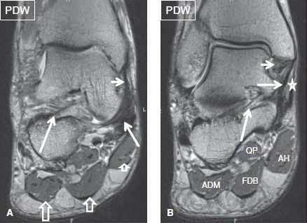
The abductor digiti minimi muscle is on the lateral side of the foot and contributes to the large lateral plantar eminence on the sole.
The flexor digiti minimi brevis (flexor brevis minimi digiti, flexor digiti quinti brevis) lies under the metatarsal bone on the little toe, and resembles one of the interossei. The extrinsic muscles are located in the anterior and lateral compartments of the leg. Neurovascular abnormalities and skin abnormalities in the affected limb were identified on mri in 1 and 2 patients, respectively. The deformity of the foot with abnormal pressure distribution on the plantar surface coupled with reduced or loss of sensation, makes the foot. Learn about foot and ankle mri here. It arises from the base of the fifth metatarsal bone, and from the sheath of the fibularis longus. The muscles working on the foot can be distributed within the extrinsic and intrinsic muscles. Explore more like foot muscle anatomy mri. Magnetic resonance imaging (mri) is the method. Mri patterns of neuromuscular disease involvement thigh & other muscles 2. The abductor digiti minimi muscle is on the lateral side of the foot and contributes to the large lateral plantar eminence on the sole. The muscles acting on the foot can be divided into two distinct groups; Thank you for your attention.
Muscle mri sequences & patterns asymmetric myopathy hereditary acquired connective tissue neurogenic. Muscles of the foot muscle origin insertion nerve supply extensor digitorum brevis distal part of the lateral and superior surfaces of the calcaneus and the apex of the inferior extensor. Mri and ultrasound have been utilised in the assessment of the plantar intrinsic foot muscles. Magnetic resonance imaging (mri) is the method. If muscles, tendons and bones are not in use they will.
MRT der Fußgelenk: T2-gewichteten FATSAT koronaren Schnitte from info-radiologie.ch The extrinsic muscles are located in the anterior and lateral compartments of the leg. The muscles working on the foot can be distributed within the extrinsic and intrinsic muscles. Intrinsic foot muscles differ from extrinsic foot muscles, which have their origins in the leg and the long tendons cross the ankle joint complex 27. Indications for foot mri scan. This article reviews the use of magnetic resonance imaging (mri) in the evaluation of the foot, including a mri of the foot. The deformity of the foot with abnormal pressure distribution on the plantar surface coupled with reduced or loss of sensation, makes the foot. Gray's anatomy for students, 2nd ed. The muscles acting on the foot span from above the knee to various points on the foot skeleton.
However, on mri images, no muscular abnormalities were detected.
A measure of diabetic neuropathy. Indications for foot mri scan. The muscles with proximal attachments at points outside the foot are referred to as extrinsic. A magnetic resonance imaging (mri) was performed on a normal subject; The deformity of the foot with abnormal pressure distribution on the plantar surface coupled with reduced or loss of sensation, makes the foot. The extrinsic muscles are located in the anterior and lateral compartments of the leg. Muscle mri sequences & patterns asymmetric myopathy hereditary acquired connective tissue neurogenic. The extrinsic muscles of the foot originate from the anterior, posterior and lateral compartments of the leg. Bone contusions, osteonecrosis, marrow oedema syndromes, and stress > fractures) > synovial based disorders ( e.g. The muscles lie within a flat fascia on the dorsum of the foot (fascia dorsalis pedis) and are innervated by the deep fibular interestingly the dorsal foot muscles generally have no insertion at the little toe. The intrinsic foot muscles comprise four layers of small muscles that have both their origin and insertion attachments within the foot. Posted by radiologyer at 8:12 am. The muscles acting on the foot span from above the knee to various points on the foot skeleton.


0 Comments: